General Surgery
We offer expert general surgery treatment for a wide range of conditions. Our board-certified surgeons specialize in treating cancer, vascular diseases, gastrointestinal issues, and breast conditions, with a focus on personalized care and advanced surgical techniques.
Expert surgical care
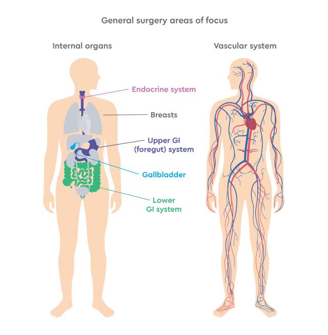
Our general surgery team provides comprehensive, compassionate care for a wide range of surgical needs. Whether you’re facing a complex vascular condition, require cancer treatment, or need care for common issues like gallbladder disease or hernias, our board-certified surgeons are here to guide you every step of the way—from diagnosis through recovery.
Our surgeons are highly trained, experienced, and committed to delivering outstanding outcomes through a patient-first approach. At SunState Medical Specialists, we use advanced technology and minimally invasive techniques whenever possible to ensure faster recovery and better overall patient experiences.
We have convenient locations in Fort Myers, Boca Raton, Destin and Niceville, FL. Through a collaborative approach with your primary care provider, we make your surgical journey as smooth and efficient as possible.

Conditions we treat
Our general surgeons treat a broad spectrum of conditions, including but not limited to:
Colorectal cancer starts in the colon or the rectum. These cancers can also be called colon cancer or rectal cancer, depending on where they start. Colon cancer and rectal cancer are often grouped together because they have many features in common.4
Stomach (gastric) cancer develops in the cells lining the stomach. The stomach is an organ on the left side of the upper abdomen that digests food. The stomach is part of the digestive tract, a series of hollow, muscular organs joined in a long, twisting tube from the mouth to the anus.5
Breast cancer is the abnormal and uncontrollable growth of the cells lining the breast lobules or ducts. These cancerous cells have the potential to spread, or metastasize, to lymph nodes near the breast or other distant parts of the body, such as the liver, lungs, bones, or brain.
Abdominal aortic aneurysm (AAA)
An abdominal aortic aneurysm is a balloon-like bulge in the aorta, the blood vessel that supplies blood from the heart to the organs. The bulge forms in the vessel wall, and if left untreated can rupture, which can be life threatening.
Carotid artery stenosis
Carotid artery stenosis is one of the leading causes of TIA and strokes. TIA is a transient ischemic attack, which has similar symptoms as a stroke but go away after a few minutes or hours and there is no brain damage. The carotid artery supplies blood to the brain and like other arteries has the ability to collect plaque, which can cause stenosis, or narrowing of the arteries, therefore allowing less blood flow to the brain.
Peripheral vascular disease (PVD)
PVD occurs when your arteries can not bring enough blood to you leg and foot muscles. Plaque build up in the arteries causes them to narrow and therefore less blood flows through to the leg. This causes pain and muscle cramping in the lower extremities after walking. In severe cases the loss of blood flow can result in gangrene and limb loss.
Gallbladder disease
Most common problems that cause pain in the gallbladder are from stones, but occasionally symptoms can arise because the gallbladder is not contracting normally. Gallstones create blockage in the gallbladder and prevent it from functioning normally.
Hernias
A hernia is a weakness or defect in the wall of the abdomen. There are several types and each is treated slightly different. The most common are inguinal and incisional. Inguinal hernias are found in the groin and may need to be repaired. Incisional hernias are found the abdomen wall, the navel, or at prior incision sites and only need to be repaired if they are painful or become large.
Gastroesophageal reflux disease (GERD)
Gastroesophageal reflux disease, or GERD, occurs when stomach acid repeatedly flows back into the esophagus, leading to heartburn. This backward flow, known as acid reflux, can irritate the esophageal lining. While occasional acid reflux is common, frequent episodes over time may develop into GERD.1
Diverticular disease
Diverticular disease refers to a group of conditions involving small pouches, called diverticula, that form in the colon. These include diverticulosis, where the pouches are present without symptoms; diverticulitis, when the pouches become inflamed or infected; and diverticular bleeding, which occurs when a blood vessel in a pouch ruptures and causes bleeding.2
Achalasia
Achalasia is a swallowing disorder that impacts the esophagus, the tube linking the mouth to the stomach. It occurs when nerve damage prevents the esophageal muscles from pushing food and liquid down into the stomach properly. As a result, food can build up in the esophagus, sometimes fermenting and coming back up into the mouth with a bitter taste.3
Thyroid disease refers to conditions that affect how the thyroid gland produces hormones, leading to either too much (hyperthyroidism) or too little (hypothyroidism) thyroxine. These imbalances can impact metabolism and cause symptoms like weight changes or irregular heartbeat. Treatment may involve medication, radiation, or surgery, depending on the condition. Thyroid nodules and goiters are also common and may need monitoring or treatment if they cause symptoms or affect hormone levels.
Parathyroid disease involves disorders where the parathyroid glands produce too much or too little parathyroid hormone (PTH), affecting calcium and phosphorus levels in the blood. Hyperparathyroidism causes high calcium and low bone density, often treated with surgery or monitored if mild. Hypoparathyroidism leads to low calcium and high phosphorus.
Breast disease
Many women develop lumps in their breasts that can be benign or cancerous. Often, benign lumps are caused by fibrocystic changes, cysts, discharge, or microcalcifications.
Request more information
Surgical procedures
Our expert general surgery team personalizes your treatment plan based on your specific condition, lifestyle, and goals. We offer a full range of general and vascular surgical options, including both minimally invasive and traditional procedures.
Gallbladder surgery
Laparoscopic surgery
Small incisions are made in the abdomen and a small camera and light are used to guide the instruments into the body and remove the gallbladder. Patients usually go home the same day.
Open surgery
A larger incision is made in the abdomen and the doctor is able to remove or opertate on the area of focus. This type of surgery has a hospital stay of 3-5 days, but in some cases is the only way to remove the gallbladder.
Diagnosis
A variety of test can be performed to determine if the gallbladder needs to be removed and the type of surgical method that should be utilized by they surgical team. An ultrasound, a CT scan, or a hepatobiliary scan (HIDA) can all be used.
Hernia repair surgery
Based on the hernia diagnosis and specific situation, either laparoscopic or open surgery is required to repair a hernia.
Inguinal surgery
The contents of the hernia are pushed back into the abdomen and the defect in the abdomen wall, or weakness, is sewn closed. Sometimes a mesh is used to reinforce the repair. This type of surgery is done as an outpatient procedure and the patient may return home shortly after the surgery.
Incisional surgery
The contents of the hernia are returned to the abdomen and the hole or weakness is sewn closed. If adjacent tissue is also weak, a synthetic mesh can be sewn in to reinforce the repair. Most of these procedures are outpatient, but in some cases a short hospital stay may be required.
Diagnosis
A physical examination can diagnose a hernia. Patients report the following symptoms:
- Bulge in the groin, abdomen, or thigh,
- Pain when lifting,
- Pain during physical activity,
- Pain while coughing or sneezing,
- Feelings of weakness or pressure in the groin and
- Discomfort when urinating
Thyroid surgery
Thyroid surgery treats conditions like goiters, hyperthyroidism, thyroid nodules, or thyroid cancer. Surgeons commonly perform hemithyroidectomy (partial removal), total thyroidectomy (full removal), and neck dissection (removal of lymph nodes). The type of surgery depends on the condition’s severity, cause, and individual needs.
Parathyroid surgery
Parathyroid surgery treats hyperparathyroidism, where overactive glands raise calcium levels, causing issues like weak bones and kidney stones. Surgeons may recommend targeted parathyroidectomy (minimally invasive), four-gland exploration (removing abnormal glands), partial parathyroidectomy (removing 3 or 3½ glands), or total parathyroidectomy (removing all glands, sometimes placing tissue in the arm).
Breast surgery
A lumpectomy or mastectomy may be used to remove the cancer, depending on the size of the lump and how far the cancer has spread. A lumpectomy only removes the lump while a mastectomy removes the entire breast. Additional treatments such as chemotherapy, radiation therapy, and hormonal therapy may be used to help destroy remaining cancer cells and reduce the risk of recurrence.
Removes the tumor and a margin (a small rim of normal breast tissue around the tumor). The breast remains intact. This is also called breast-conserving surgery.
For small tumors, limited breast surgery has been shown to have similar outcomes to mastectomy when additional treatments such as radiation therapy are added.
A mastectomy is a more extensive surgery that involves the removal of the entire breast tissue, including the skin, and sometimes the nipple and areola. This type of surgery is often recommended when the tumor is large relative to the breast size or if there are multiple tumors within the breast.
Diagnosis
If you feel a change in your breast, it is best to see a doctor and have a mammogram. An ultrasound of the breast may also be used to determine if a lump is solid or filled with fluid. If needed a biopsy of the lump is performed.
- Non-Surgical Biopsy: fine needle aspiration, ultrasound-guided, and stereotactic biopsies are all performed without anesthesia. A sample of the lump is removed and tested to determine if the lump is benign or cancerous.
- Surgical Biopsy: a surgical biopsy allows a larger sample to be obtained. This may require wire localization. A small, thin wire is placed in the breast to mark the tissue that is to be removed. In the operating room the wire and the lump is removed and the mass is tested.
Foregut surgery
Our expert team of general surgeons, nurses, and staff specializes in the diagnosis and surgical treatment of foregut conditions, which affect:
- esophagas
- stomach
- upper small inestines (duodenum and jejunum)
We offer both conventional and minimally invasive foregut procedures, using the latest technology to reduce recovery time, minimize scarring, and improve outcomes.
Every treatment plan is personalized to the patient’s specific symptoms and needs. If you're considering surgery for a foregut condition, our skilled surgeons are here to provide compassionate, comprehensive care tailored to each patient.
Colon surgery
The colon is removed during surgery and the intestine is sewn back together. The procedure generally requires a short hospital stay of about five days.
Colectomy
Colectomy is a surgical procedure to remove all or part of your colon. Your colon, part of your large intestine, is a long tubelike organ at the end of your digestive tract. Colectomy may be necessary to treat or prevent diseases and conditions that affect your colon.6
Hemicolectomy
Hemicolectomy is a surgical procedure that involves the removal of a portion of the colon, either the left or right side, to treat various conditions such as colon cancer, inflammatory bowel disease, or diverticulitis. The remaining healthy sections of the colon are then reconnected to restore bowel function.7
Diagnosis
Colon health can be assessed using various diagnostic tools, including colonoscopy to visually inspect the colon and collect tissue samples, biopsies to analyze suspicious tissue for cancer, and blood tests to evaluate overall health and monitor tumor markers like CEA.8
Abdominal aortic aneurysm (AAA) repair
Small aneurysms can be safely observed. Larger aneurysms may require open or endovascular surgery, depending on the size and shape of an individual’s blood vessels.
Open surgery
An incision is made in the abdomen. An artificial graft is inserted inside the damaged artery. Recovery time is longer (5-8 days in the hospital) than endovascular repairs, but for many patients it is the only option to treat an AAA.
Endovascular surgery
Two small incisions are made in the groin and the artificial graft is inserted into the artery. This procedure is less invasive than open surgery and recovery time is shorter with patients usually going home 1-2 days after surgery.
Diagnosis
Usually there are no symptoms and an aneurysm is often found during a routine exam for an unrelated problem. Anyone is at risk to develop an abdominal aortic aneurysm, however, individuals that smoke, have high blood pressure, are over 55(men) or 65(women), or have a close relative with an AAA is at greater risk.
Routine examinations, ultrasounds of the abdomen, and an abdominal CT scan can help diagnosis an abdominal aortic aneurysm.
Peripheral vasuclar disease (PVD) bypass surgery
While the best way to manage Peripheral Vascular Disease (PVD) is by reducing your risk factors and preventing its development, medication may be used in some cases—under your doctor’s supervision—to help relieve claudication (pain when walking).
For more advanced stages of PVD, bypass surgery is often the primary treatment option. There are two main types of bypass procedures:
Femoral popliteal bypass surgery
The blocked artery is bypassed via the popliteal artery in the upper part of the leg using either an artificial graft or you own vein.
Distal bypass surgery
The blocked artery is bypassed via the popliteal artery in the upper part of the leg using either an artificial graft or you own vein.
Endovascular surgery
Endovascular surgery is a less invasive procedure using balloons and stents that does not require anesthesia. It aids in wound healing and pain with claudication. This surgery may be used in less severe cases of PVD.
Diagnosis
Peripheral Vascular Disease can be diagnosed using several different tests, including the ultrasound Doppler test, ankle brachial index (ABI), and angiograms. Patient reported symptoms include:
- Claudication (cramping pain in the buttocks, thigh, or calf when exercising and relieved when resting)
- rest pain (constant pain in the foot due to restriction of blood flow)
- nonhealing ulcers on the foot or toes
- gangrene
Risk factors include smoking, hypertension, diabetes mellitus and family history of PVD
Carotid artery stenosis surgery
There are two ways to treat carotid artery stenosis, by carotid endarterectomy or by carotid artery angioplasty and stenting. Your surgeon will decide the best treatment options based on the patient's diagnostic results.
Endarterectomy
This is an open surgery used to remove plaque from the artery. It is very effective and most patients return home within 24 hours of the operation.
Angioplasty and stenting
This less invasive procedure is used to improve the blood flow through the carotid artery. A balloon is used to expand the narrowed artery and then a stent is placed in the artery. The stent is left in place to hold the artery open, and therefore allow normal blood flow to the carotid artery.
Diagnosis
Carotid bruits are sounds that are heard by using a stethoscope on the neck that can indicate a build up of plaque in the artery. A carotid duplex scan can detect the degree of stenosis. An MRA is often used to confirm the diagnosis. It is a non invasive test that uses magnetic resonance imagining.
In addition a contrast angiogram is used as a diagnostic procedure and prior to carotid angioplasty and stenting. It requires a catheter in the groin to inject dye into the artery. Then x-rays are obtained to identify where and to what degree the artery is blocked by plaque, allowing the surgeon to select the most suitable treatment option for the patient.